Based on the cited paper, it is clear that [18F]-FDOPA Positron Emission Tomography/ Computed Tomography (PET/CT) plays a crucial role in the follow-up of Glioblastoma Multiforme (GBM). This is particularly important when conventional imaging methods like Magnetic Resonance Imaging (MRI) yield inconclusive results, often in the challenging differentiation between tumor recurrence and radiation necrosis.
[18F]-FDOPA is an excellent PET imaging radiotracer for both primary and metastatic brain lesions due to its high tumor-to-normal-brain background ratio. This superior contrast allows for better visualization of tumor extent compared to regions with normal brain tissue. The mechanism behind this enhanced uptake in brain tumors is attributed to the over-expression of the amino-acid transporter LAT-1 in tumor cells and their vasculature. This increased expression eventually leads to a higher accumulation of [18F]-FDOPA within the tumor compared to the surrounding normal brain tissue.
The literature cited in this paper indicates that [18F]-FDOPA PET/CT demonstrates higher diagnostic accuracy than [18F]-FDG PET/CT in detecting primary or recurrent gliomas. This is significant because [18F]-FDG uptake can be limited in brain tumors due to its significant uptake in normal brain tissue, potentially masking subtle tumor activity making the interpretation of results more difficult.
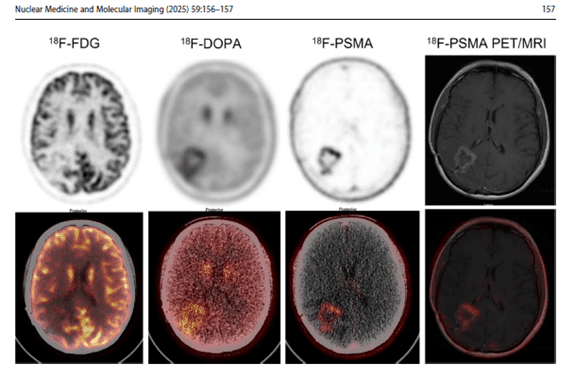
The “Interesting Images” section in the paper serves as a compelling case study illustrating the importance of [18F]-FDOPA in GBM follow-up. In this case, a 42-year-old man with recurrent GBM presented with an enhancing lesion on MRI that could have represented either tumor recurrence or radiation necrosis. While [18F]-FDG PET/CT showed only a photopenic area with equivocal metabolic activity, both [18F]-FDOPA and [18F]-PSMA-1007 PET/CT clearly demonstrated a focal area of increased tracer uptake in the right parietal lobe, corresponding to the enhanced region appearing on MRI. This increased uptake on [18F]-FDOPA PET/CT was indicative of an active tumor characterized by increased amino acid metabolism. The post-hoc fusion images, overlaying the PET data on the MRI images, further highlighted the advantage of these PET tracers in identifying the active tumor.
This case underscores the value of incorporating [18F]-FDOPA as an adjunctive method for evaluating recurrent gliomas, especially in difficult cases where MRI results are ambiguous. Moreover, it emphasizes the importance of a multimodal imaging strategy that integrates conventional MRI with advanced PET techniques like [18F]-FDOPA PET/CT.
In conclusion, the evidence presented in this paper strongly suggests that [18F]-FDOPA PET/CT is a vital tool in the monitoring of GBM patients. Its high tumor-to-background ratio, linked to the overexpression of amino acid transporters in tumor cells, provides superior diagnostic accuracy compared to [18F]-FDG PET/CT in detecting recurrent disease. The case study effectively demonstrates its utility in resolving ambiguous MRI findings, particularly in distinguishing tumor recurrence from radiation necrosis. Therefore, [18F]-FDOPA PET/CT plays a significant role in the comprehensive evaluation and management of recurrent GBM.
Reference
- Usmani, S., Marafi, F., Sadeq, A. et al. Spectrum of Metabolic and Angiogenic Imaging with 18F-FDOPA and 18F-PSMA 1007 PET/CT in the Evaluation of Recurrent Glioblastoma Multiforme. Nucl Med Mol Imaging59, 156–157 (2025).
https://doi.org/10.1007/s13139-024-00895-w

Haim Golan
MD MSc
Chief Medical Officer
Medical Adviser
Isotopia Molecular Imaging LTD


