Prostate cancer continues to pose a significant health challenge, impacting 1 in 8 men in the United States [1]. Early and accurate diagnosis is critical, as it can mean the difference between successful treatment and advanced disease progression. Thus, there is a pressing need for advanced diagnostic tools that can reliably identify cancerous lesions.
Among the advancements in nuclear medicine, PSMA-11 has emerged as a groundbreaking tool in prostate cancer imaging, redefining standards and offering unparalleled diagnostic accuracy [2].
Understanding PSMA-11
Prostate-Specific Membrane Antigen (PSMA) is an overexpressed cell surface protein in prostate cancer cells, including both primary and metastatic lesions.
PSMA-11 is a Gallium-68 radiolabeled ligand that binds specifically to this antigen, allowing for precise imaging of prostate cancer tissues using PET-CT. The development and clinical adoption of PSMA-11 has been driven by its high binding affinity to PSMA, which significantly enhances the sensitivity and specificity of cancer detection.
The specificity of PSMA-11 for PSMA receptors means the images produced are highly accurate, thereby reducing the risk of false positives and enabling precise localization of cancerous tissues.
Clinical Advantages of PSMA-11
One of the primary reasons PSMA-11 is considered the gold standard is its exceptional diagnostic accuracy.
For instance, research has demonstrated that 68Ga-PSMA-11 imaging significantly reduces the incidence of false positives.
In matched pair comparisons, 68Ga-PSMA-11 has been shown to produce up to 5 times fewer false positives compared to 18F-PSMA-1007 [3].
This reduction in false positives is crucial for accurate diagnosis and effective treatment planning, as it minimizes the risk of unnecessary interventions and ensures that treatment decisions are based on reliable data [3].
Scientific Validation and Clinical Evidence
The clinical efficacy of PSMA-11 has been validated through numerous studies and clinical trials. Key research has highlighted the tracer’s effectiveness in various aspects of prostate cancer imaging:
- Rauscher et al. [3] demonstrated the superiority of 68Ga-PSMA over 18F-PSMA-1007 in reducing false positives and enhancing diagnostic accuracy. This study provided strong evidence supporting the use of PSMA-11 as a preferred tracer for prostate cancer imaging.
- Alberts et al. [4] directly compared the clinical performance and cost efficacy of 68Ga-PSMA-11 and 18F-PSMA-1007, in diagnosing recurrent prostate cancer. 244 patient’s imaging results were analyzed using a Markov chain decision model to evaluate clinical outcomes and cost data. 18F-PSMA-1007 exhibited a higher PET positivity rate but also a higher rate of uncertain and false-positive findings compared to 68Ga-PSMA-11. The conclusion was that 68Ga-PSMA-11 demonstrated superior clinical performance and cost efficacy in most jurisdictions analyzed.
These studies, among others [5], have established PSMA-11 as the benchmark for prostate cancer imaging, reflecting its advanced performance and clinical utility.
Conclusion
In summary, PSMA-11 has set a new standard in prostate cancer imaging with its high binding affinity, reduced false positives, and superior image quality. As a high-affinity ligand for PSMA, PSMA-11 enables precise PET imaging, offering critical advantages in the diagnosis and management of prostate cancer. The extensive clinical validation and evidence supporting its efficacy underscore its position as the gold standard in the field.
For professionals in nuclear medicine, the adoption of PSMA-11 represents a significant advancement in diagnostic capabilities.
By integrating PSMA-11 into their clinical practice, practitioners can enhance the accuracy of prostate cancer imaging, improve patient outcomes, and contribute to the ongoing advancement of oncology diagnostics.
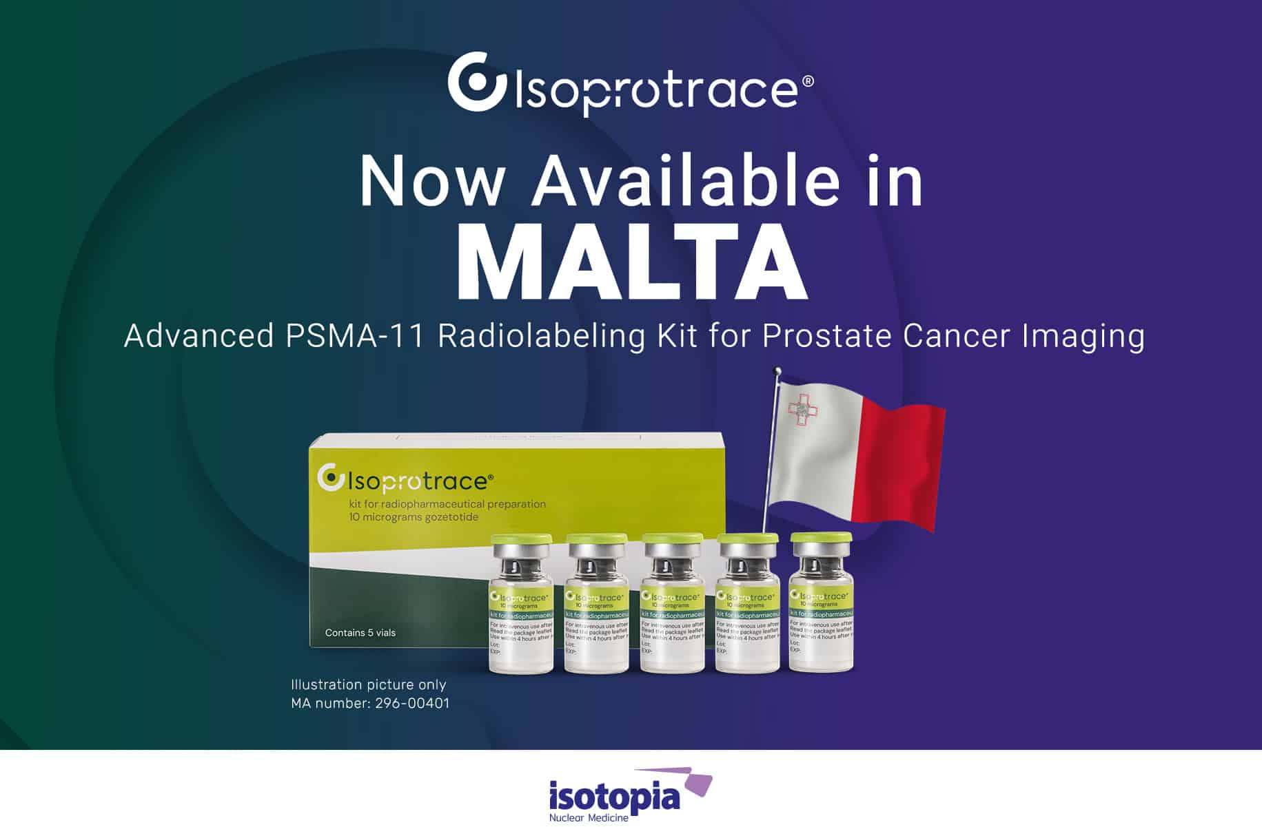
Isoprotrace®: PSMA-11 Radiolabeling Kit for Prostate Cancer Imaging
Isoprotrace®, developed by Isotopia™, is a PSMA-11 radiolabeling kit tailored for prostate cancer imaging.
This ready-to-use, multi-dose kit facilitates the efficient preparation of Gallium-68 (68Ga) Gozetotide solution for injection. Specifically indicated for use in Positron Emission Tomography (PET), Isoprotrace® is designed to detect PSMA-positive lesions in men with prostate cancer.
References:
- American Cancer Society. https://www.cancer.org/cancer/types/prostate-cancer/about/key-statistics.html
- Hofman, Michael S. et al. Prostate-specific membrane antigen PET-CT in patients with high-risk prostate cancer before curative-intent surgery or radiotherapy (proPSMA): a prospective, randomised, multicentre study. The Lancet, Volume 395, Issue 10231, 1208 – 1216
- Rauscher, I. et al. (2020). Comparison of 68Ga-PSMA-11 and 18F-PSMA-1007 for Prostate Cancer Imaging. Journal of Nuclear Medicine, 2020 Jan; 61(1):51-57.
- Alberts I, Mingels C, Zacho HD, Lanz S, Schöder H, Rominger A, Zwahlen M, Afshar-Oromieh A. Comparing the clinical performance and cost efficacy of [68Ga]Ga-PSMA-11 and [18F]PSMA-1007 in the diagnosis of recurrent prostate cancer: a Markov chain decision analysis. Eur J Nucl Med Mol Imaging. 2022.
- Purysko AS, Abreu AL, Lin DW, Punnen S Not All Prostate-Specific Membrane Antigen Imaging Agents Are Created Equal: Diagnostic Accuracy of Ga-68 PSMA-11 PET/CT for Initial and Recurrent Prostate Cancer. Appl Radiol. 2024;53(2):22-35

Haim Golan
MD MSc
Chief Medical Officer
Medical Adviser
Isotopia Molecular Imaging LTD


